![]() Figure 1 of
Palamalai, Mol Vis 2006;
12:1543-1551.
Figure 1 of
Palamalai, Mol Vis 2006;
12:1543-1551.
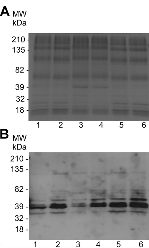
Figure 1. Nitrotyrosine western analyses of rod outer segment proteins from rats exposed to intense light
A representative Coomassie blue-stained gel (A) and corresponding western blot (B) of rod outer segment (ROS) proteins with antinitrotyrosine antibody is shown. Lane 1: animals unexposed to light with dimethylthiourea (DMTU) treatment; lane 2: unexposed to light without DMTU; lane 3: exposed to light for 8 h with DMTU treatment; lane 4: exposed to light for 8 h without DMTU treatment; lane 5: 24 h after 8 h of light exposure with DMTU treatment; lane 6: 24 h after 8 h of light exposure without DMTU. Quantification of nitrotyrosine in each lane is represented graphically as mean±standard deviation (C). Results are from three gels from three independent sets of ROS. In each case, the unexposed DMTU untreated nitrotyrosine optical density was set to 100%.