![]() Figure 3 of
Ayala-Ramirez, Mol Vis 2006;
12:1483-1489.
Figure 3 of
Ayala-Ramirez, Mol Vis 2006;
12:1483-1489.
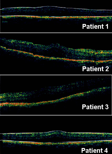
Figure 3. Optical coherence tomography images in patients
Optical coherence tomography images in left eye of patients 1 and 2 and right eye of patients 3 and 4. Diffuse macular thickening, outer retinal layer schisis with discrete bridging elements (foveoschisis) were evident in all affected subjects.