![]() Figure 1 of
Ayala-Ramirez, Mol Vis 2006;
12:1483-1489.
Figure 1 of
Ayala-Ramirez, Mol Vis 2006;
12:1483-1489.
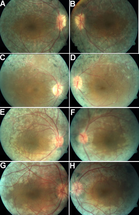
Figure 1. Fundus photographs of patients
Fundus photographs of patient number 1 (propositus, A, B), and affected siblings patient number 2 (C, D), number 3 (E, F), and number 4 (G, H). Left column represents right eye (OD); right column represents left eye (OS). Optic disc drusen, pigment clumping, bone-spicule pigmentation, blunting of the macular reflex and minimal vascular attenuation are evident.