![]() Figure 5 of
Vieira, Mol Vis 2006;
12:1448-1460.
Figure 5 of
Vieira, Mol Vis 2006;
12:1448-1460.
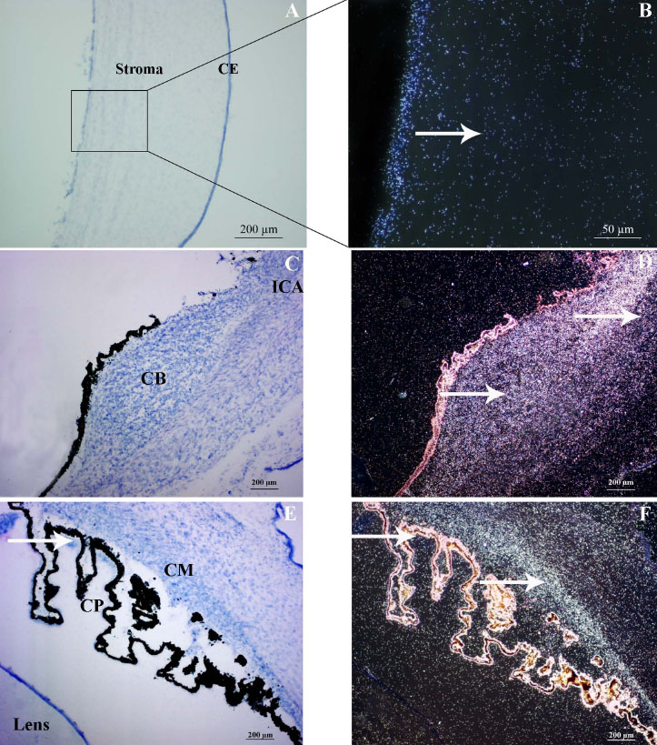
Figure 5. PITX2 gene expression at fifteen weeks of gestation in fetal ocular tissue sections
On the left side Bright-field (A, C, and E) and in the right side the corresponding Dark Field aspects (B, D, and F) of PITX2 gene expression at fifteen weeks of gestation. The specific signals are shown by white arrows (B, D, and F). PITX2 mRNAs were detected in corneal endothelium (CE) and stroma (A and B). PITX2 mRNAs were also strongly detected in irido-corneal angle (ICA; C and D) and in the ciliary body especially in the non pigmented cell layer of the ciliary processes (CP) and in the developing ciliary muscles (CM; E and F). Autofluorescence is shown in the pigmented cell layer of the ciliary processes (D).