![]() Figure 6 of
Burgess, Mol Vis 2006;
12:1437-1447.
Figure 6 of
Burgess, Mol Vis 2006;
12:1437-1447.
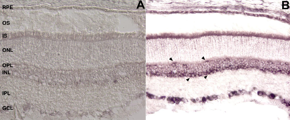
Figure 6. In situ hybridization staining for MFG-E8 in the rat eye
In situ hybridization staining of rat eye with sense (A) and antisense (B) dig-labeled 503 base RNA probes to MFG-E8. The RPE, photoreceptor cell inner segments (IS) and some ganglion cells (GCL) are heavily stained. Cells on the inner and outer layers of the INL also show staining (arrowheads). Outer segments are present in (B) but show no staining with the riboprobe. Original magnification equals to 40x.