![]() Figure 4 of
Burgess, Mol Vis 2006;
12:1437-1447.
Figure 4 of
Burgess, Mol Vis 2006;
12:1437-1447.
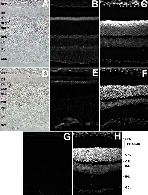
Figure 4. Immunofluorescence staining of MFG-E8 in the mouse eye
Immunofluorescence staining of MFG-E8 in mouse eyes using antibodies 204AP (A, B, C), and 18A2 (D, E, F). A represents brightfield, B shows 204AP staining, C shows DAPI staining of the same section. In the absence of primary antibody, no staining was observed (not shown). D is brightfield, E is 18A2 staining, and F is DAPI staining of the same section. G shows stainins with no primary antibody and H is DAPI staining of G. In both sections shown, the POS layer has torn with the result that a thin layer of POS is attached to the apical surface of the RPE. Original magnification = 40x.