![]() Figure 3 of
Burgess, Mol Vis 2006;
12:1437-1447.
Figure 3 of
Burgess, Mol Vis 2006;
12:1437-1447.
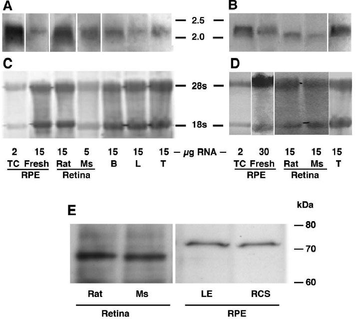
Figure 3. MFG-E8 mRNA and protein in rat tissues
A, B: Northern blots showing the presence of MFG-E8S (A) and MFG-E8L (B) in rat and mouse tissues. C, D: Methylene Blue stained transblots showing the loading and integrity of the total RNA. Each of the stained lanes in C and D corresponds to the lane above it in A and B. Note that different amounts of total RNA were loaded in each lane. Figures (A), (B), (C) and (D) are composite figures assembled from several separate gels, each run with kbp standards to distinguish between the long and short transcripts. The dig-labeled 207 base long probe (B) shows some reactivity with MFG-E8S mRNA, resulting in short form mRNA showing in rat and mouse retina and cultured RPE in (B). E: Western blot showing the presence of MFG-E8S in extracts of rat and mouse retina, and fresh RPE from LE and RCS rats. TC represents tissue culture; Ms represents mouse; T represents testis, B represents brain, L represents liver.