![]() Figure 6 of
Ricard, Mol Vis 2006;
12:1427-1436.
Figure 6 of
Ricard, Mol Vis 2006;
12:1427-1436.
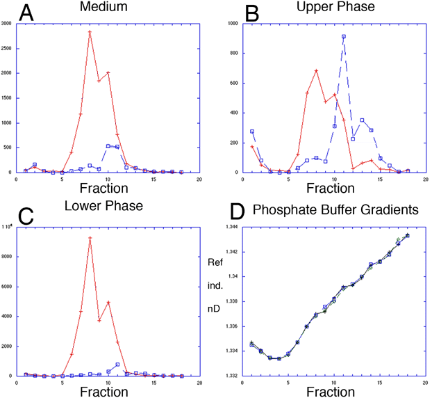
Figure 6.
Myocilin protein was modified by palmitic acid. Hydroxylapatite chromatograms of phase-separated myocilin. Protein labeled with 35S (dashed blue squares) and 3H palmitic acid (solid red crosses) was immunoprecipitated, treated with SDS, reduced and heated, then adsorbed to HAP columns and eluted with 0.1 to 0.5 M phosphate gradient. Y-axis=dpm; X-axis=fraction. A: Myocilin from culture medium resolved in two peaks, each containing palmitate and protein label. B: The upper phase from Triton X-114 resolved as three or more peaks, each containing palmitate and protein label. C: The lower phase from Triton X-114 resolved as one lipid and one protein-lipid peak. D: Y-axis=refractive indices (nD) measured for fractions eluted from HAP chromatography and showing identical elution gradients for samples in A (blue squares), B (black crosses), and C (green, triangles).