![]() Figure 4 of
Ricard, Mol Vis 2006;
12:1427-1436.
Figure 4 of
Ricard, Mol Vis 2006;
12:1427-1436.
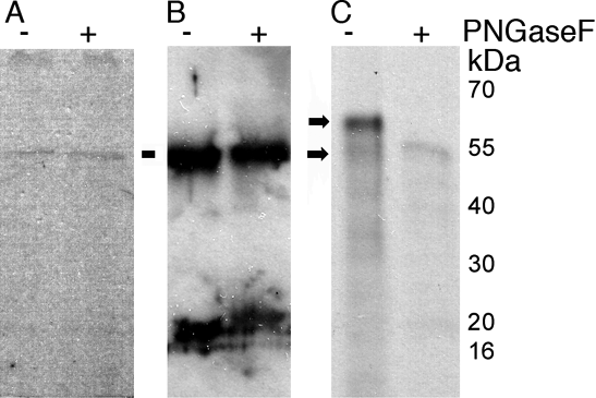
Figure 4.
Myocilin was N-glycosylated in MDCK. 35S labeled extracellular myocilin protein was immunoprecipitated from the medium of canine cells, and a portion was treated with peptide endoglycanase F (PNGase F). A: Fluorography of untreated and treated passage 2 beagle ONH cell myocilin. B: Western blot of beagle ONH cell myocilin. C: Fluorography of MDCK myocilin. Untreated (upper arrow) and an approximately 54 kDa protein backbone remained after treatment (lower arrow). Immunoprecipitates were separated by electrophoresis on 4-20% gradient SDS gels, and visualized by fluorography on x-ray film. The western blot was visualized with chemiluminescence.