![]() Figure 2 of
Ricard, Mol Vis 2006;
12:1427-1436.
Figure 2 of
Ricard, Mol Vis 2006;
12:1427-1436.
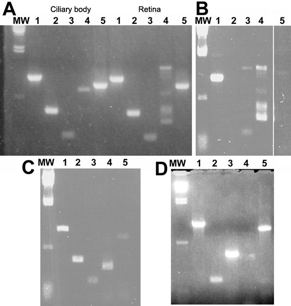
Figure 2.
Gene expression in canine ocular cells. RT-PCR was performed on RNA extracted from tissue or cultured cells, using primers that are listed in Table 2. Primers were designed to cross exon boundaries to rule out genomic DNA amplification. A: Ciliary body (left), and retina (right). Lane 1, myocilin; lane 2, Rab 3 GDI; lane 3, NCAM; lane 4, GFAP; lane 5, β-actin. B: Optic nerve head. Lane 1, myocilin; lane 2, Rab 3 GDI; lane 3, NCAM; lane 4, GFAP; lane 5, β-actin. C: Cells cultured from optic nerve head. Lane 1, myocilin; lane 2, NCAM; lane 3, GDI; lane 4, GFAP; lane 5, β-actin. D: MDCK cells. Lane 1, myocilin; lane 2, GFAP; lane 3, NCAM; lane 4, GDI; lane 5, β-actin.