![]() Figure 9 of
Dvoriantchikova, Mol Vis 2006;
12:1417-1426.
Figure 9 of
Dvoriantchikova, Mol Vis 2006;
12:1417-1426.
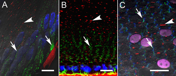
Figure 9.
Double immunostaining for Panx1 and Cx50 in P5 mouse lenses. No significant colocalization of punctuate labeling specific to Panx1 (green) and Cx50 (red) was detected in either sagittal (A) or equatorial lens sections (B and C). Connexin gap junction plaques are indicated by arrowheads and pannexin aggregates by arrows. Nuclei were stained with DAPI (blue in A and B or magenta in C). F-phalloidin staining for actin in C appears in blue. The anti-Cx50 antibody had some cross-reactivity with lens capsule that also appears in red (A and B). The scale bar represents 10 mm.