![]() Figure 8 of
Dvoriantchikova, Mol Vis 2006;
12:1417-1426.
Figure 8 of
Dvoriantchikova, Mol Vis 2006;
12:1417-1426.
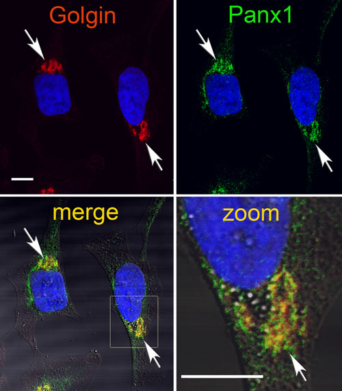
Figure 8.
Immunostaining of HeLa cells for the Golgin-97 (red) and endogenous Panx1 (green). Panx1-specific labeling was localized to the perinuclear region and colocalized (yellow) extensively with Golgi-specific labeling (arrows). The boxed region is shown enlarged in A (zoom). DAPI staining for nuclei is shown in blue. The scale bar represents 10 mm.