![]() Figure 7 of
Dvoriantchikova, Mol Vis 2006;
12:1417-1426.
Figure 7 of
Dvoriantchikova, Mol Vis 2006;
12:1417-1426.
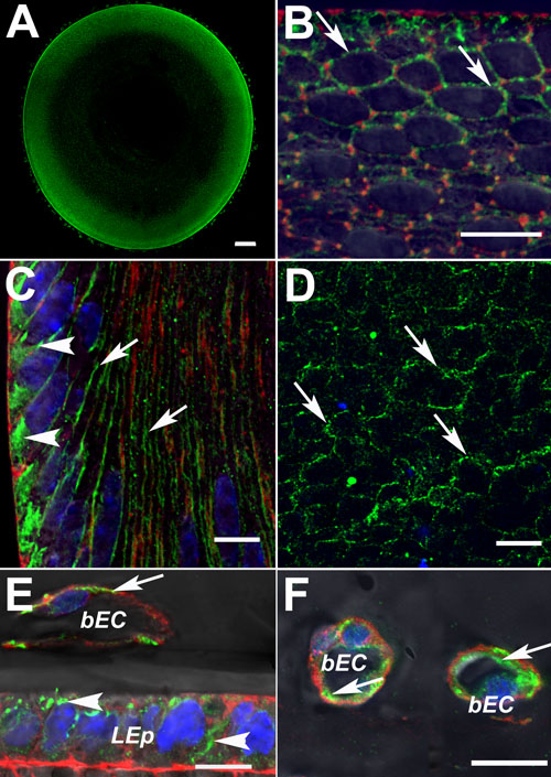
Figure 7.
Immunohistochemistry for Panx1 in mouse lenses. In equatorial lens slices, Panx1-specific labeling (green) was evident in the epithelium and young elongating fibers (A, B). In young elongating fibers (B, C), lens epithelial cells (E), and blood endothelial cells of tunica vasculosa lentis (E, F), Panx1-specific labeling was observed in the perinuclear region (arrowheads) and as punctuate staining in plasma membranes (arrows). Mature fibers (D) showed only punctuate staining in plasma membranes. F-phalloidin staining for actin is shown in red (B,C,E,F); DAPI staining for nuclei is shown in blue (C,D,E,F); bEC designates blood endothelial cells and LEp designates lens epithelium. The scale bar represents 50 mm in A and 10 mm in B-F.