![]() Figure 4 of
Dvoriantchikova, Mol Vis 2006;
12:1417-1426.
Figure 4 of
Dvoriantchikova, Mol Vis 2006;
12:1417-1426.
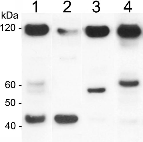
Figure 4.
Fractionation of Panx1 isoforms by differential centrifugation. Three Panx1 isoforms, 43, 58, and 62 kDa, were quantitatively separated using this approach. Lens homogenate fractionated by differential centrifugation and separated by SDS-PAGE was probed with anti-Panx1 antibodies. Lanes: 1, total lens homogenate; 2, water-soluble proteins (100,000 g supernatant); 3, water insoluble proteins pelleted at 7,000 g; 4, microsomal, organelle-enriched fraction pelleted by spinning the supernatant from lane 3 at 100,000 g.