![]() Figure 6 of
Bernstein, Mol Vis 2006;
12:147-155.
Figure 6 of
Bernstein, Mol Vis 2006;
12:147-155.
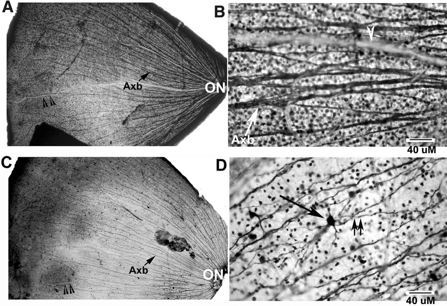
Figure 6. Distribution of Bex-ir in rat retina following optic nerve stroke
Bex-ir immunoreactivity in control (A) and rAION-affected (C) flat mounted retinae. Antibody to Bex1 was reacted against whole retina, developed using DAB-immunohistochemistry, and examined at low and high magnification. A: Control and quarter of the retina flat mounted, low magnification. Axon fibers are distributed evenly and densely within bundles (Axb) to supply the entire retina. Bex positive cell bodies are visible between axons. B: Control retina flat mount, high magnification. Both Bex-ir positive axons and cell bodies are present. A blood vessel is also visible (white arrowhead). C: One-quarter of a retina flat mount 21 days post-rAION. Retinal ganglion cell axonal loss is visible as a decreased density of axon fiber bundles. D: Bex-ir in rAION retina, higher magnification. There is decreased axon fiber density, and decreased density of Bex-ir labeled cells. Large Bex-ir positive cells (Large black arrow) with long processes typical of axons (double arrows) are present in the stroke-affected tissue, which are not readily seen in the control section (arrows). The optic nerve (ON) is identified. The scale bars represent 40 μm.