![]() Figure 4 of
Bernstein, Mol Vis 2006;
12:147-155.
Figure 4 of
Bernstein, Mol Vis 2006;
12:147-155.
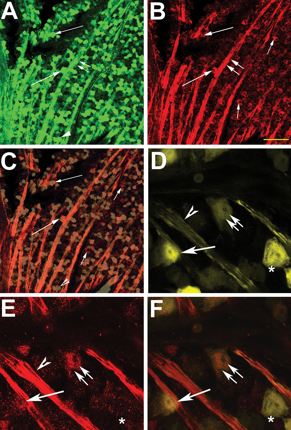
Figure 4. Co-expression of Bex-immunoreactivity and cyan fluorescent protein in in retinal ganglion cell axons and cell bodies of transgenic mice
Bex1 antibody was reacted with fixed whole retinas from Thy-1 CFP expressing transgenic mice. A: CFP expression in axons and RGCs. CFP protein is strongly expressed in both RGC axons (double arrows) and RGC cell bodies of different sizes (single arrow). B: Bex-ir localization. Bex-ir positive cells are visible (long arrows), and strong expression present in axon bundles (double arrows). Some Bex-positive cells do not express CFP in the confocal image (short arrows). C: Confocal (merged) image. Bex-ir and CFP co-localization are visible (long arrow). Isolated Bex-ir is evident in a few cells (short arrows). A few isolated CFP positive (Bex-ir negative) cells are also seen (arrowhead). D-F: CFP and Bex expression at high magnification. D: Retinal CFP expression. Strong CFP signal is apparent in soma (arrows) and axon bundles (arrowhead), with reduced CFP expression in other cells (double arrows). One strongly CFP positive (+) cell has minimal Bex-ir (asterisk). E: Bex-ir in the same section. Bex-ir is strongest in axons, while variable expression is seen in RGC soma. One CFP positive cell is not apparent with Bex-ir (asterisk). F: Merged confocal image. Both CFP and Bex-ir are present in individual cells, suggesting that RGCs are expressing Bex-ir. The asterisk identifies a CFP(+)/Bex-ir cell. The scale bar represents 70 μm in A-C and 25 μm in D-F.