![]() Figure 1 of
Bernstein, Mol Vis 2006;
12:147-155.
Figure 1 of
Bernstein, Mol Vis 2006;
12:147-155.
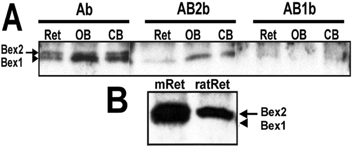
Figure 1. Characterization of Bex1 and Bex2 expression in mouse and rat central nervous system and retinal tissues
A: Extracts of retina (Ret; 5 mg), olfactory bulb (OB; 0.5 mg), and cerebellum (CB; 0.5 mg) from postnatal day 14 (P14) mice were subjected to immunoprecipation with the Bex1 antiserum followed by electrophoresis on SDS-PAGE, blotting to nitrocellulose and probing with anti-Bex1 antiserum before (Ab) and after pre-adsorption with Bex1 (AB1b) or Bex2 protein (AB2b). This analysis demonstrated the presence of Bex1 (arrowhead) and Bex2 (arrow) proteins in the mouse retina when using Ab. When antibody preabsorbed with recombinant Bex2 protein (AB2b) was used, only Bex1 protein (arrowhead) was detected. B: Rat and mouse retinas express high levels of Bex proteins. Immunoprecipitated extracts of adult mouse (mRet) or rat (ratRet) retinas (5 mg each) were analyzed by immunoblotting with anti-Bex1 antiserum (Ab). Mouse retina gave strong signals for both Bex1 and Bex2. By contrast, rat retina apparently expresses only Bex2.