![]() Figure 2 of
Lyu, Mol Vis 2006;
12:1403-1410.
Figure 2 of
Lyu, Mol Vis 2006;
12:1403-1410.
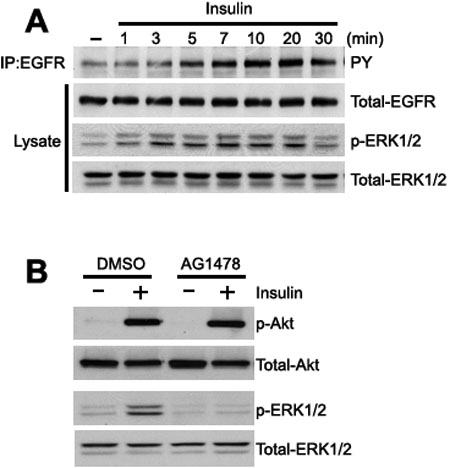
Figure 2.
Insulin induces EGFR phosphorylation in THCE cells. A: Serum- and growth-factor-starved THCE cells were incubated with or without 5 μg/ml insulin for various times and lysed. Cell lysates were immunoprecipitated with EGFR antibody and the immune complexes subjected to immunoblotting with phosphotyrosine (PY20H) antibody (top). Blots were reprobed with EGFR antibody as an equal loading control. The lysates were fractionated by 10% SDS-PAGE and immunoblotted with phospho-ERK1/2 or ERK1/2 antibodies (bottom). B: To inhibit EGFR activity, we added 1 μM AG1478 to serum- and growth-factor-starved THCE cells for 1 h. The cells were further incubated with or without 5 μg/ml insulin for 10 min, lysed, and immunoblotted with phospho-Akt or phospho-ERK1/2 antibody. Total Akt or ERK/12 was probed to normalize protein loading.