![]() Figure 1 of
Lyu, Mol Vis 2006;
12:1403-1410.
Figure 1 of
Lyu, Mol Vis 2006;
12:1403-1410.
Figure 1.
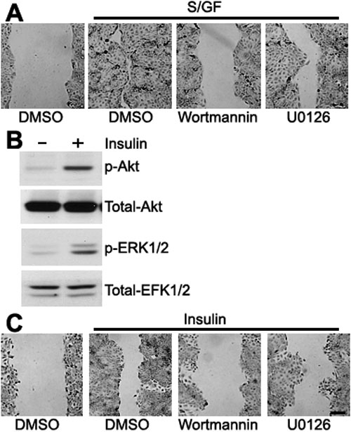
Insulin stimulates wound healing by activating PI3K and ERK. A: A wound was introduced into a monolayer of THCE cells with a micropipette tip, and closure of the wound was examined in cultures incubated for 12 h in medium containing serum and growth factors, or vehicle. Next, 100 nM wortmannin, 10 μM U0126, or DMSO were added to the THCE cells for 1 h before scratch wounding. B: THCE cells starved of serum and growth factors were incubated with 5 μg/ml insulin for 10 min and lysed 10 min later. The lysates were immunoblotted with antiphospho-Akt, and antiphospho-ERK1/2, as well as with anti-Akt or anti-ERK1/2 to normalize protein loading. C: Serum- and growth-factor-starved THCE cells were pretreated with 100 nM wortmannin, 20 μM U0126, or DMSO, wounded by scratching and incubated with 5 μg/ml insulin for 12 h. Each experiment was performed at least three times. Scale bar, 200 μm.