![]() Figure 6 of
Liang, Mol Vis 2006;
12:1392-1402.
Figure 6 of
Liang, Mol Vis 2006;
12:1392-1402.
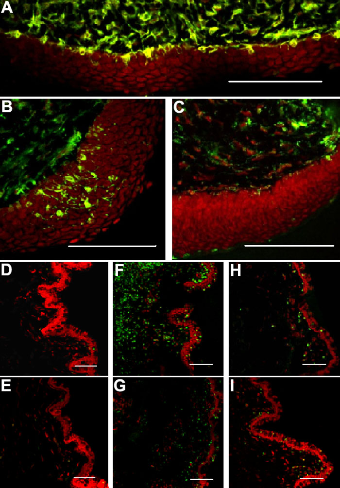
Figure 6.
A, B, and C: Immunofluorescence staining of vimentin (green) and propidium iodide (red) showing dendritiform cells. Subconjunctival LPS administration increased dendritiform cell density at the junction of the conjunctival epithelium and the substantia propria (A), and also within the conjunctival epithelium (B). C: Subconjunctival BSS injections showed weak infiltration of vimentin-positive cells. D-I: Immunofluorescence staining of CD4 (D, F, and H) or CD8 (E, G, and I) in conjunctivas injected with BSS (D and E), LPS (F and G), or LPS+anti-TNF-α (H and I). The scale bars indicate 100 μm.