![]() Figure 5 of
Liang, Mol Vis 2006;
12:1392-1402.
Figure 5 of
Liang, Mol Vis 2006;
12:1392-1402.
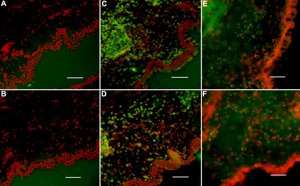
Figure 5.
Immunofluorescence staining of TNF-α (A,C,E) and TNFR-1 (B,D,F) in conjunctiva (A,B) injected with BSS, injected with LPS (C,D), or injected with LPS+anti-TNF-α (E,F). Immunostaining of TNF-α and TNFR-1 appears in green and nuclei are in red following propidium iodide staining. The scale bar indicates 100 μm.
