![]() Figure 3 of
Liang, Mol Vis 2006;
12:1392-1402.
Figure 3 of
Liang, Mol Vis 2006;
12:1392-1402.
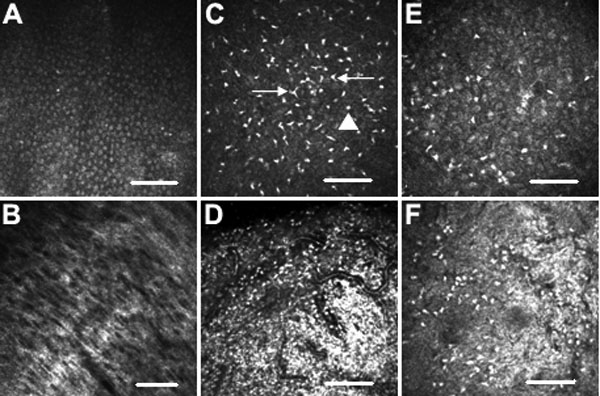
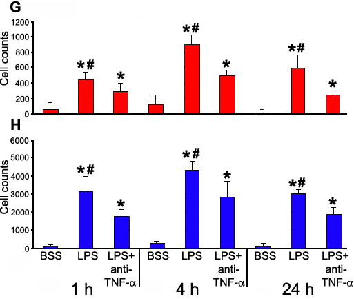
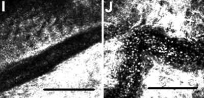
Figure 3.
Heidelberg Retina Tomograph II in vivo confocal microscopy images of conjunctival epithelium (A, C, and E) and substantia propria (B, D, and F) 4 h after injections of BSS (A and B), LPS (C and D), and LPS+anti-TNF-α (E and F; 400x400 μm). C: The dendritic-shaped cells (arrows) were surrounded by many smaller round cells suggestive of lymphocytes (arrowheads). Cell counts of inflammatory cell infiltration in the epithelium/mm2 (G) and substantia propria/mm2 (H). The asterisk indicates a p<0.001 compared to BSS-injected conjunctivas and the sharp (hash mark) denotes a p<0.01 compared to LPS+anti-TNF-α-injected conjunctivas. Images of a blood vessel in a BSS-injected conjunctiva I and in a LPS-injected conjunctiva (J) show in vivo rolling and leukocyte migration of inflammatory cells. The scale bar indicates 100 μm.