![]() Figure 1 of
Liang, Mol Vis 2006;
12:1392-1402.
Figure 1 of
Liang, Mol Vis 2006;
12:1392-1402.
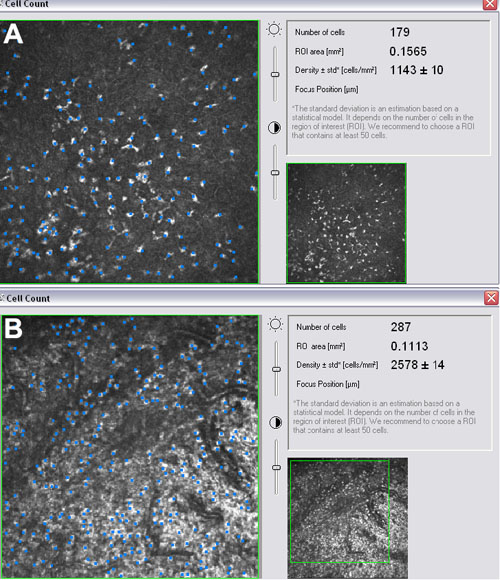
Figure 1.
Inflammatory cell infiltrations were analyzed separately in the conjunctival epithelium (A) or substantia propria (B) near the injection area with Cell Count® software in manual mode. The data were expressed as density ±SD (cells/mm2).