![]() Figure 4 of
Singh, Mol Vis 2006;
12:1372-1379.
Figure 4 of
Singh, Mol Vis 2006;
12:1372-1379.
Figure 4.
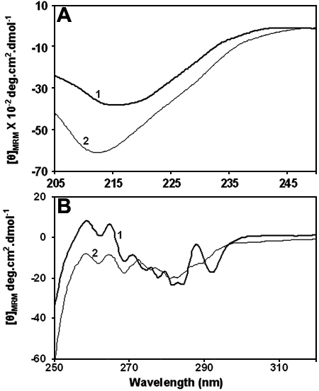
Circular dichroism (CD) spectra of wild type and G98R αA-crystallin. A: Far-UV CD spectra of the wild type (curve 1) and the mutant (curve 2) αA-crystallin indicating a significant change in the secondary structure of G98R αA-crystallin upon mutation. B: Near-UV CD spectra of the wild type (curve 1) and the mutant αA-crystallin (curve 2) indicating a loosening of the tertiary structural packing of G98R αA-crystallin upon mutation. [θ]MRM, mean residue mass ellipticity.
