![]() Figure 2 of
Sun, Mol Vis 2006;
12:1364-1371.
Figure 2 of
Sun, Mol Vis 2006;
12:1364-1371.
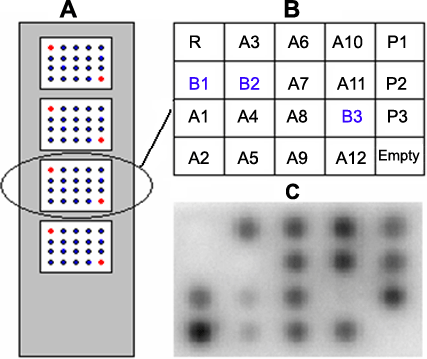
Figure 2.
Analysis of microarray hybridization of selected ASO probes against survivin. A: Microarray sketch map. B: Schematic representation of a microarray. Table 1 shows the A, B, P, and R representing oligonucleotides. C: Hybridization image of 32P-labeled transcripts to microarrays of oligonucleotides.