![]() Figure 4 of
Chen, Mol Vis 2006;
12:1355-1363.
Figure 4 of
Chen, Mol Vis 2006;
12:1355-1363.
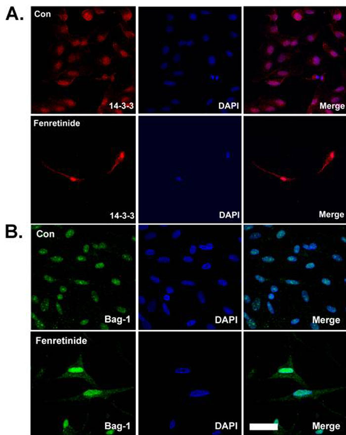
Figure 4. Distribution of 14-3-3 and bag-1 proteins in ARPE-19 cells treated with fenretinide
ARPE-19 cells were treated with 1.0 μM fenretinide for 5 days and analyzed by confocal microscopy. Cells were stained with A: anti 14-3-3 antibody and Cy3 conjugated secondary antibody; B: anti bag-1 antibody and Alexa Fluor 488 conjugated secondary antibody. Cell nuclei were stained with DAPI (blue). Scale bar represents 50 μm.