![]() Figure 2 of
Hammer, Mol Vis 2006;
12:1348-1354.
Figure 2 of
Hammer, Mol Vis 2006;
12:1348-1354.
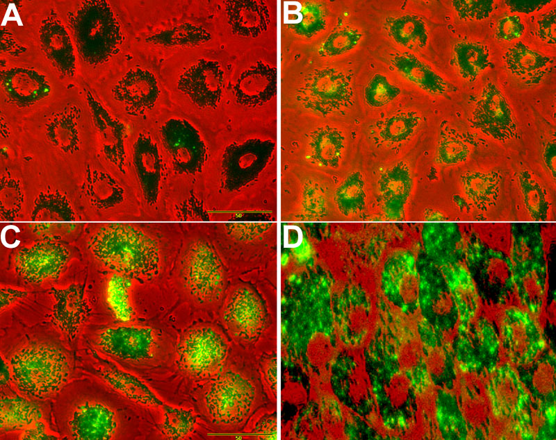
Figure 2. A2-E supplementation to retinal pigment epithelium cells
Red channel represents phase contrast image; green channel represents auto-fluorescence. A: Cells grown in the absence of A2-E (control). B: Cells after incubation with 10 μM A2-E for 24 h. C: Cells after incubation with 10 μM A2-E for 48 h. D: Cells 72 h after withdrawal of A2-E supplemented for 48 h.