![]() Figure 3 of
Andrieu-Soler, Mol Vis 2006;
12:1334-1347.
Figure 3 of
Andrieu-Soler, Mol Vis 2006;
12:1334-1347.
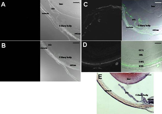
Figure 3. Adult mice eye sections one hour after transcorneosceral iontophoresis of CY5-labeled ribozymes
Confocal microscopy of anterior segment in phase contrast and fluorescence one hour after saline iontophoresis (A). Confocal microscopy of anterior segment in fluorescence and phase contrast one hour after application of fluorescent ribozymes without current (B). Confocal microscopy of anterior segment in fluorescence and phase contrast one hour after application of fluorescent ribozymes with current (C). Confocal microscopy of posterior segment in fluorescence and phase contrast one hour after application of fluorescent ribozymes with current (D). Hematoxylin-eosin stained sections one hour after application of fluorescent ribozymes with current (E). Scale bars represents 100 μm.