![]() Figure 2 of
Andrieu-Soler, Mol Vis 2006;
12:1334-1347.
Figure 2 of
Andrieu-Soler, Mol Vis 2006;
12:1334-1347.
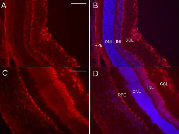
Figure 2. Retinal distribution of Hex-labeled oligonucletides
In vivo light-controlled distribution of Hex-labeled oligonucletides in the retina after intravitreous injection of vectosomes in the rat eye. Fluorescence microphotographs of retinal sections 24 h after intravitreous injections of vectosomes without illumination (A) and (B): with Dapi nuclei co-staining, or with illumination (C) and (D): with Dapi nuclei co-staining. Retinal pigment epithelium (RPE), outer nuclear layer (ONL), inner nuclear layer (INL), and ganglion cell layer (GCL). Scale bars represents 100 μm.