![]() Figure 2 of
Aldave, Mol Vis 2006;
12:142-146.
Figure 2 of
Aldave, Mol Vis 2006;
12:142-146.
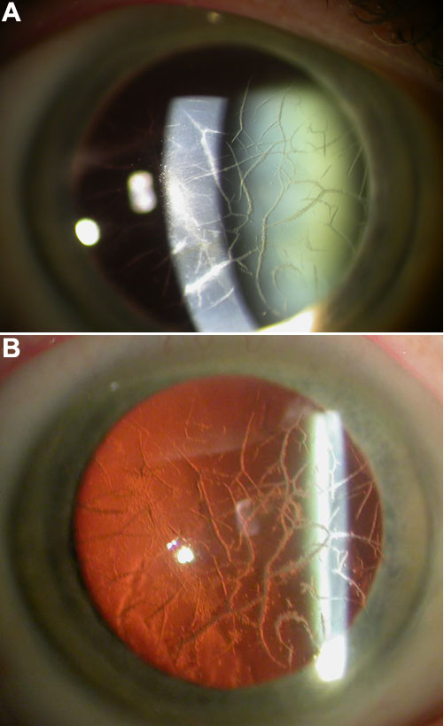
Figure 2. Unilateral lattice corneal dystrophy
Shown in this slit lamp photomicrograph of the right cornea are thin, branching, diffusely distributed anterior stromal lattice lines, seen with both direct (A) and indirect (B) illumination. This pattern is consistent with the corneal changes in lattice corneal dystrophy type 1.