![]() Figure 7 of
Tserentsoodol, Mol Vis 2006;
12:1319-33.
Figure 7 of
Tserentsoodol, Mol Vis 2006;
12:1319-33.
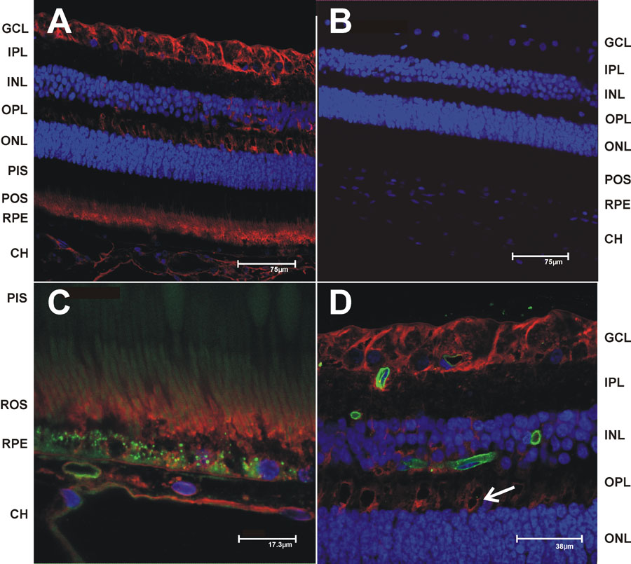
Figure 7. Immunohistochemical localization of lecithin:cholesterol acyltransferase in monkey retina
The vibrotome sections from monkey retina were processed for immunhistochemistry and imaged by fluorescent confocal microscopy (see Materials and Methods). Nuclei were stained with DAPI (blue) and immunoreactivity was detected using a Cy5 conjugated secondary antibodies (red). A: Lecithin:cholesterol acyltransferase (LCAT) was localized using a rabbit polyclonal to a recombinant C-terminus peptide (Abcam Inc. Cambridge, MA) at 1:1000. B: No primary antibody control. C: LCAT immunoreactivity at higher magnification focusing on the photoreceptors and retinal pigment epithelium/choriocapillaris regions. D: LCAT immunoreactivity at higher magnification focusing on the OPL and GCL regions. Primer pairs used for the RT-PCR analyses of the different genes shown in Fig. 1. The GenBank accession numbers for the cDNAs from which the primers were selected are shown in the right side column. The products generated were sequenced to confirm their authenticity. Images C and D are shown with the green channel to take advantage of the retinal autofluorescence and provide better structural definition. Capillaries in D were stained with isolectin IB4 (green). Scale bars were included with each image.