![]() Figure 4 of
Tserentsoodol, Mol Vis 2006;
12:1319-33.
Figure 4 of
Tserentsoodol, Mol Vis 2006;
12:1319-33.
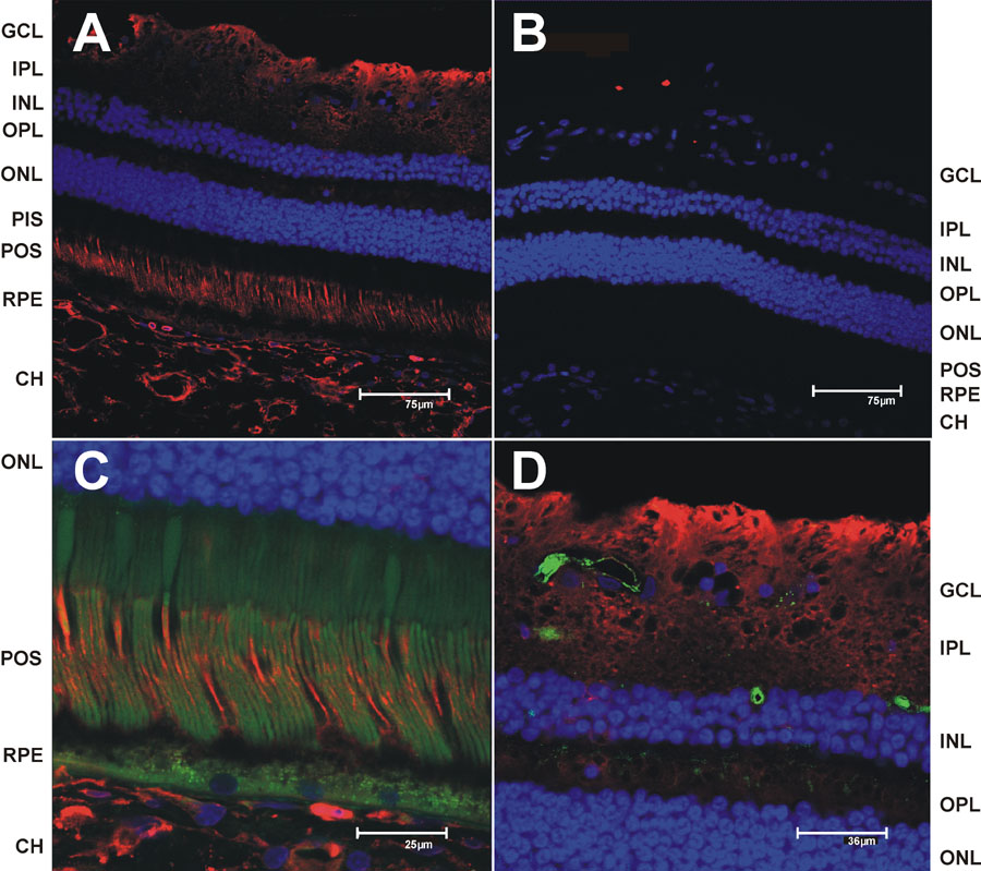
Figure 4. Immunohistochemical localization of SR-BI in monkey retina
The vibrotome sections from monkey retina were processed for immunhistochemistry and imaged by fluorescent confocal microscopy (see Materials and Methods). Nuclei were stained with DAPI (blue) and immunoreactivity was detected using a Cy5 conjugated secondary antibodies (red). A: SR-BI immunoreactivity detected using anti SR-BI peptide SPAAKGTVLQEAKL (cross-reacts with many species) rabbit polyclonal antibody (Abcam Inc.) at 1:100 dilution. B: No primary antibody control. C: SR-BI immunoreactivity at higher magnification focusing on the photoreceptors and retinal pigment epithelium/choriocapillaris regions. D: SR-BI immunoreactivity at higher magnification focusing on the GCL regions. Images C and D are shown with the green channel to take advantage of the retinal autofluorescence and provide better structural definition. Images C and D are shown with the green channel to take advantage of the retinal autofluorescence and provide better structural definition. Capillaries were in D were stained with isolectin IB4 (green). Scale bars were included with each image.