![]() Figure 3 of
Tserentsoodol, Mol Vis 2006;
12:1306-1318.
Figure 3 of
Tserentsoodol, Mol Vis 2006;
12:1306-1318.
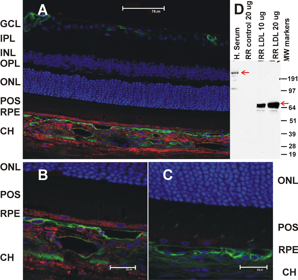
Figure 3. Immunohistochemical localization of human apoB in the rat retina 4 h after human low density lipoprotein injection
Vibrotome sections of rat retina 4 h post-injection with human CTL-LDL were imaged by confocal microscopy after incubation with anti-human apoB (1:200). The anti-human apoB immunoreactivity was detected using a Alexa Fluor® 633-conjugated donkey anti-sheep secondary antibody (red; see Materials and Methods). The nuclei were stained with DAPI (blue) and capillaries were stained with Alexa 488®-conjugated isolectin IB4 (green). A: Low magnification of the rat retina demonstrating human apoB immunoreactivity in the choriocapillaris (CH) and RPE. B: Higher magnification of the CH and RPE regions. C: Control retina (no LDL injection). D: Immunoblot of CTL-LDL injected and untreated rat retina (RR) demonstrating the 70 kDa (lower arrow) processed form of human apoB. Human serum was used a positive control for apoB (upper arrow).