![]() Figure 1 of
Lim, Mol Vis 2006;
12:1302-1305.
Figure 1 of
Lim, Mol Vis 2006;
12:1302-1305.
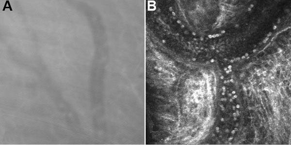
Figure 1.
Non-enhanced, representative images of ocular vessels obtained from A the ASL-1000 white light scanning confocal microscope (Advanced Scanning Ltd, New Orleans, LA) and B the HRTII laser confocal microscope (Heidelberg Engineering, Germany). Magnification of both images was 400X.