![]() Figure 4 of
Wu, Mol Vis 2006;
12:1292-1302.
Figure 4 of
Wu, Mol Vis 2006;
12:1292-1302.
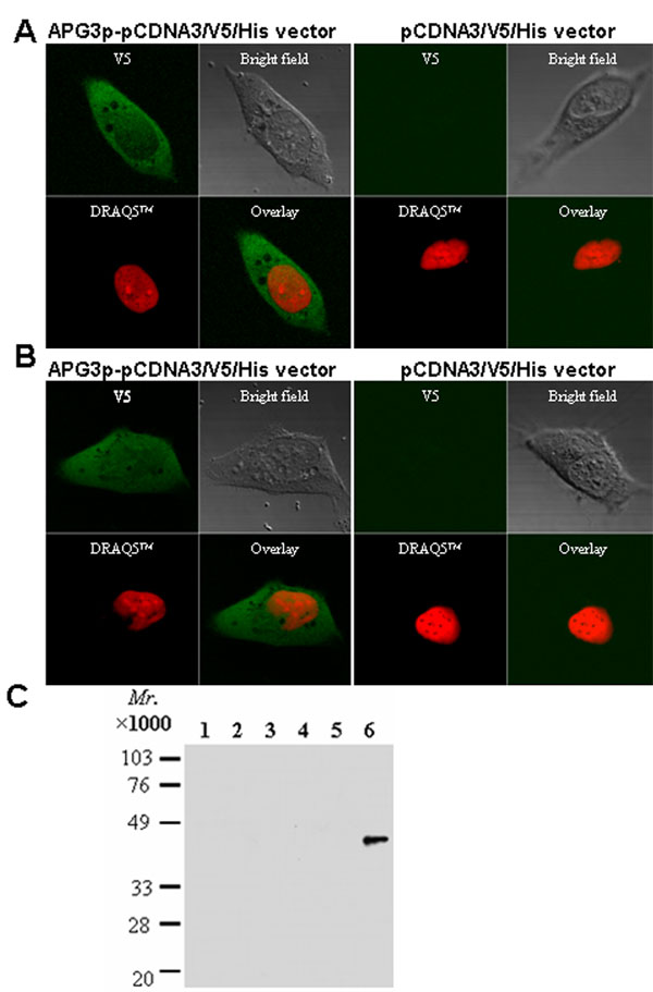
Figure 4. Expression, immunocytochemistry and subcellular fractionation
Plasmid-expressing rat Apg3p and pCDNA3/V5/His vector were used to transfect the chinese hamster ovary (CHO, A) and a transformed mouse kidney cell line (COS-7; B) cells. Forty eight h after transfection, the cells were treated with DRAQ5TM for 10 min for nucleic staining and then fixed, permeabilized, and stained with the anti-V5 antibody followed by an FITC-conjugated secondary antibody. (C): Untransfected (lanes 1-3) and Apg3-transfected (lanes 4-6) CHO cells were fractionated by differential centrifugation, and the fractions were blotted with the anti-His-tag antibody. Lanes 1 and 4, insoluble pellet after low speed centrifugation; 2 and 5, microsomal proteins; 3 and 6, soluble cytosolic proteins.