![]() Figure 2 of
Wu, Mol Vis 2006;
12:1292-1302.
Figure 2 of
Wu, Mol Vis 2006;
12:1292-1302.
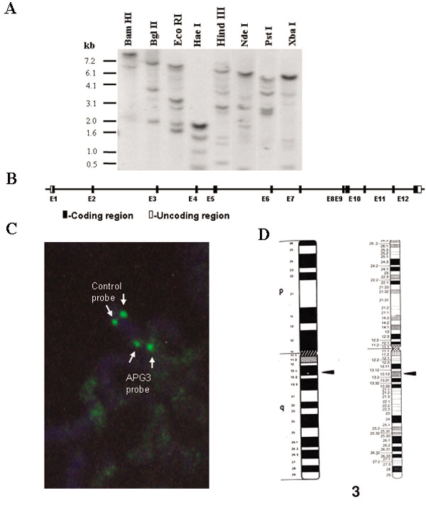
Figure 2. Genomic Southern blot analysis of rat Apg3L gene and the structure and chromosomal location of human APG3L gene
A: Ten μg of genomic DNA from Sprague-Dawley rats were separately digested with restriction enzymes as described in Methods, followed by electrophoresis and Southern blot analysis using a full-length rat Apg3 cDNA as a probe. B: Schematic representation of the human APG3L gene: black and open boxes represent coding exons and untranslated regions, respectively; introns are represented by the connecting lines. C: Fluorescent in situ hybridization of human metaphase chromosomes. Arrows indicate binding of a human APG3L genomic DNA clone and a control clone previously mapped to 3q26, both labeled with dUTP. D: Ideogram with the relative positions of the human APG3L gene localized to chromosome 3q13.1 as indicated by arrowheads.