![]() Figure 1 of
Riazuddin, Mol Vis 2006;
12:1283-1291.
Figure 1 of
Riazuddin, Mol Vis 2006;
12:1283-1291.
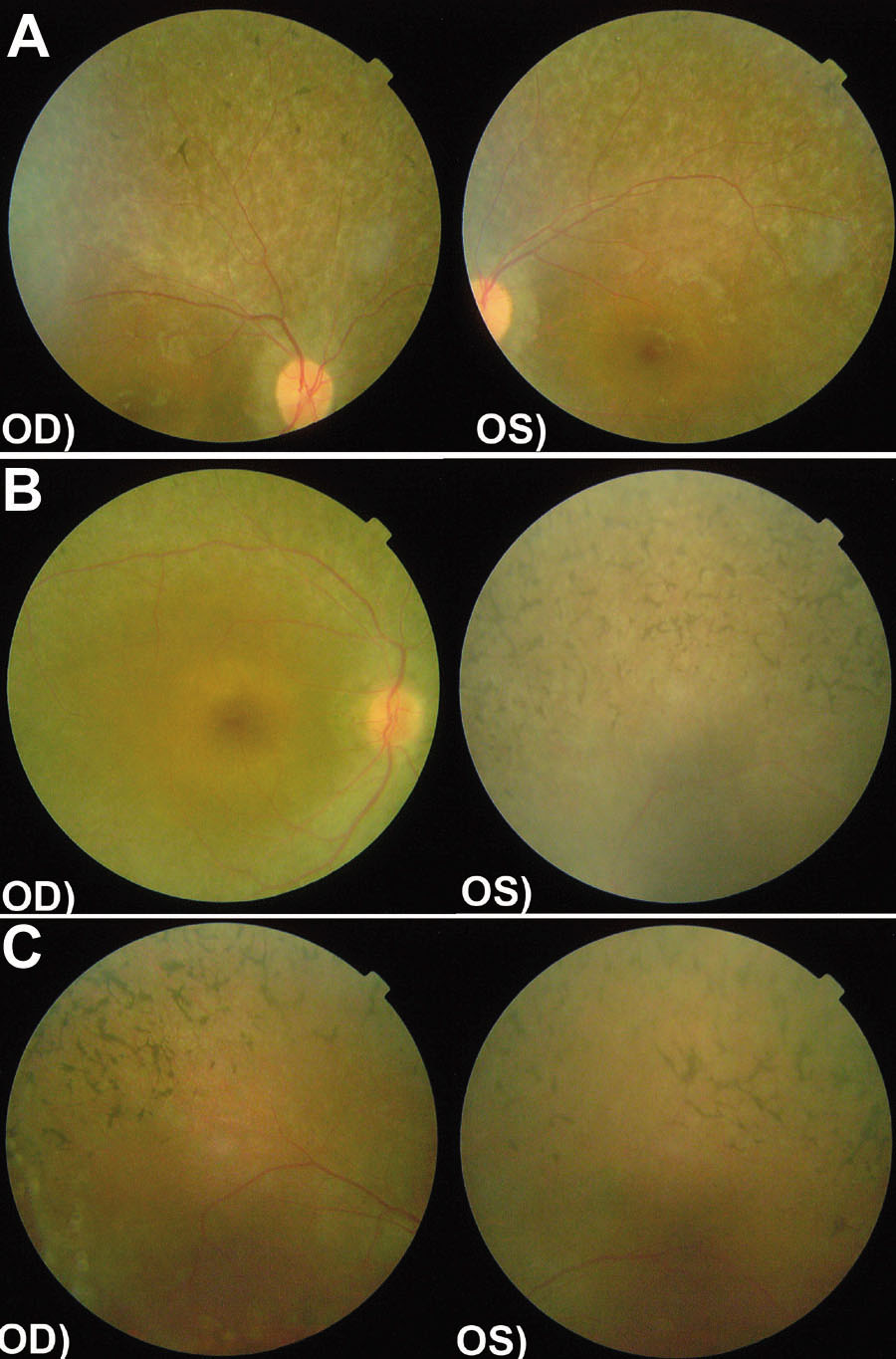
Figure 1. Fundus photograps of affected individuals
Funduscopy photographs of (A) individual 20 of family 61019 (affected 18 year old, B) individual 12 of family 61021 (affected 33 year old) and (C) individual 10 of family 61074 (affected 25 year old). There are changes typical of retinitis pigmentosa including attenuation of retinal arteries and bone-spicule pigment deposits in the mid periphery of the retina.