![]() Figure 2 of
Pelzel, Mol Vis 2006;
12:1272-1282.
Figure 2 of
Pelzel, Mol Vis 2006;
12:1272-1282.
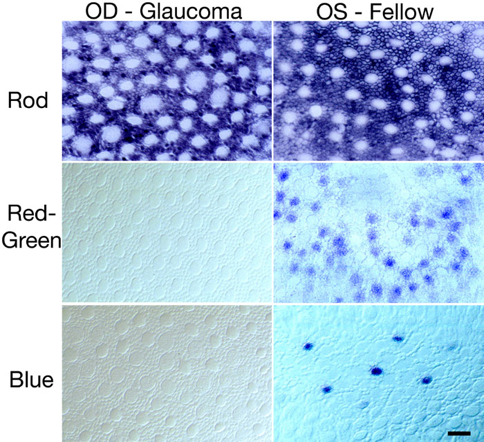
Figure 2. In situ hybridization of opsin probes in experimental glaucoma
Tissue samples from both eyes were taken from the inferotemporal mid-peripheral retina (see text). Stained tissue was embedded in glycol methacrylate and sectioned through the inner segment layer to visualize opsin localization. Sections were viewed using Nomarski interference microscopy. The control retina shows a normal pattern of rod, red/green, and blue cone opsin. The glaucomatous retina shows swollen cone inner segments and no detectable staining for either of the cone probes. The rod opsin staining is similar to that of the control retina. The scale bar represents 10 μm.