![]() Figure 1 of
Pelzel, Mol Vis 2006;
12:1272-1282.
Figure 1 of
Pelzel, Mol Vis 2006;
12:1272-1282.
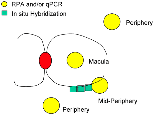
Figure 1. Map of harvested retina tissues
Samples were harvested from the posterior poles of fresh monkey eyes using a 3 mm trephine (yellow circles) for RNA analysis (RNase protection assays represents RPA, and quantitative real time PCR represents qPCR), or dissected from fixed eyes for in situ hybridization studies (green boxes). Red oval represents the optic disc. The major retinal arteries are shown. The mid-periphery region shown in this schematic is one of the regions of earliest detectable damage in humans with glaucoma. Peripheral segments of retina were taken at different locations, but principally in the far superotemporal or inferior regions near the ora serrata. Both regions of peripheral retina yielded similar results in our experiments (data not shown).