![]() Figure 7 of
Hu, Mol Vis 2006;
12:1250-1258.
Figure 7 of
Hu, Mol Vis 2006;
12:1250-1258.
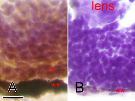
Figure 7. Cryosection and immunostaining of the wildtype and Egr1 morphants
Cryosections of zebrafish eyes at 72 h postfertilization with zpr-1 immunostaining for photoreceptor cells. The wildtype (A) has markedly more labeled retinal cells in the outer nuclear layer (*) than the Grade-3 Egr1 morphant (B). The retinal pigmented epithelial layer (**) is also much thinner in the morphant. The scale bar represents 10 μm in photo A, and is applicable to photo B.