![]() Figure 6 of
Hu, Mol Vis 2006;
12:1250-1258.
Figure 6 of
Hu, Mol Vis 2006;
12:1250-1258.
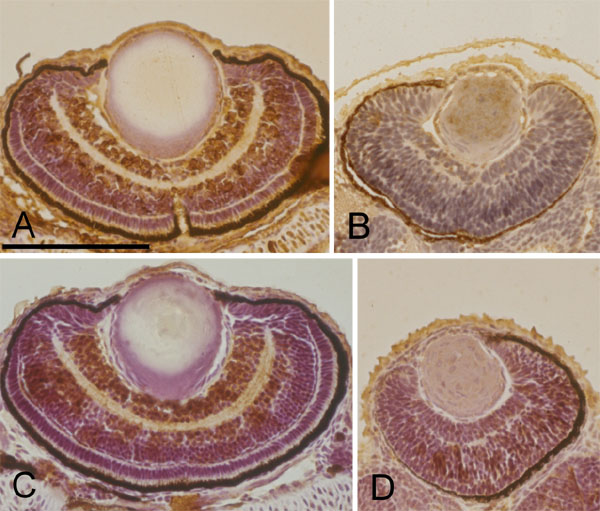
Figure 6. Immunohistochemical staining of the wildtype and Egr1 morphants
Horizontal sections of zebrafish eyes at 72 h postfertilization with immunohistochemical stain for glutamate receptor 1 (A, B) and acetylated α-tubulin (C, D). Compared with the wildtype (A, C), retinal cells of the Grade-3 Egr1 morphant (B, D) arranged more compactly and disorderly. Significantly smaller areas of staining for both glutamate receptor 1 and acetylated α-tubulin appear in the morphant's retina. Scale bar represents 100 μm in panel A, and is applicable to all panels.