![]() Figure 4 of
Jo, Mol Vis 2006;
12:1243-1249.
Figure 4 of
Jo, Mol Vis 2006;
12:1243-1249.
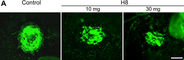
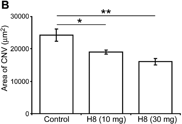
Figure 4.
H8 suppressed the development of CNV lesions at sites of laser induced rupture of Bruch's membrane. Choroidal flat-mounts were examined by fluorescence microscopy following immunostaining with PECAM-1 to identify vessels. A: Animals treated with control antibody showed large areas of CNV as compared to animals treated with either 10 or 30 mg/kg of H8. B: Quantitation of CNV lesion size in IgG and H8 treated samples. Both 10 mg/kg and 30 mg/kg treatments caused a statistically significant reduction in lesion size as compared to control untreated animals (the asterisk indicates a p<0.05 and the double asterisk denotes a p<0.001). Scale bar=20 μm.