![]() Figure 3 of
Jo, Mol Vis 2006;
12:1243-1249.
Figure 3 of
Jo, Mol Vis 2006;
12:1243-1249.
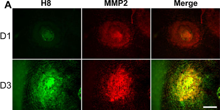
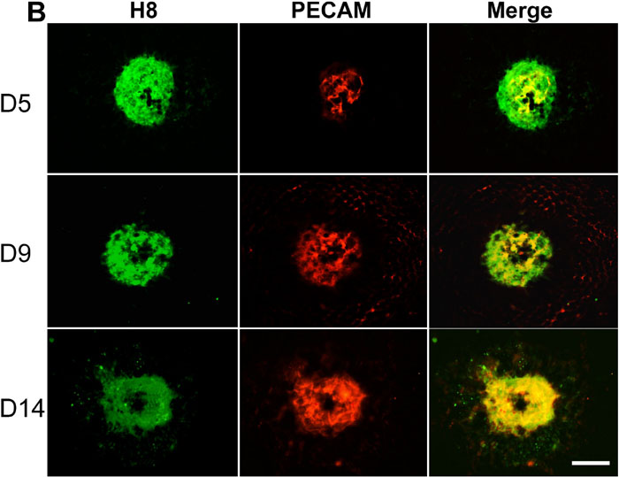
Figure 3.
Time-course of expression of collagen type IV cryptic epitopes following Bruch's membrane rupture by laser photocoagulation. A: Early CNV samples were costained with H8 (green) and MMP-2 (red) at days 1 and 3 following injury (D1, D3). MMP-2 staining precedes H8 at D1 and colocalizes with denatured collagen IV at D3. Scale bar=80 μm. B: Choroidal flat-mounts obtained at 5, 9, and 14 days (D5, D9, D14) after laser injury and costained with H8 (green) and PECAM-1 (red). H8 staining precedes neovessel growth at day 5 (D5) and colocalizes with neovessels by day 14 (D14). C: Choroidal flat mounts obtained at day 9 (D9) after laser injury were costained with H8 (green) and a collagen type IV antibody (red). Collagen type IV staining of the CNV lesion marked a larger area than did H8 staining. Scale bar=40 μm.