![]() Figure 1 of
Jo, Mol Vis 2006;
12:1243-1249.
Figure 1 of
Jo, Mol Vis 2006;
12:1243-1249.
Figure 1.
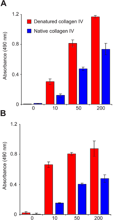
H8 binds to denatured collagen IV. A: ELISA showing that H8 preferentially recognizes heat denatured collagen IV. B: Binding specificity of HUIV26, the antibody from which H8 was derived. C: The cryptic collagen type IV site recognized by H8 is revealed by MMP-2 induced proteolysis. Murine retinal sections were incubated for 2 h with either control buffer or MMP-2 (1.0 μg/ml). Subsequently, sections were stained with H8 (green) and lectin (red). H8 staining in control (arrowhead) and MMP-2 treated samples (arrow). In the image, INL identifies the inner nuclear layer of the retina, ONL marks the outer nuclear layer of the retina; and CH indicates the choroid. The scale bar in the H8, Lectin, and Merge panels is equal to 50 μm. Human IgG (green) was used as an isotype-matched irrelevant antibody control and the scale bar is equal to 100 μm

