![]() Figure 3 of
Taliana, Mol Vis 2006;
12:1233-1242.
Figure 3 of
Taliana, Mol Vis 2006;
12:1233-1242.
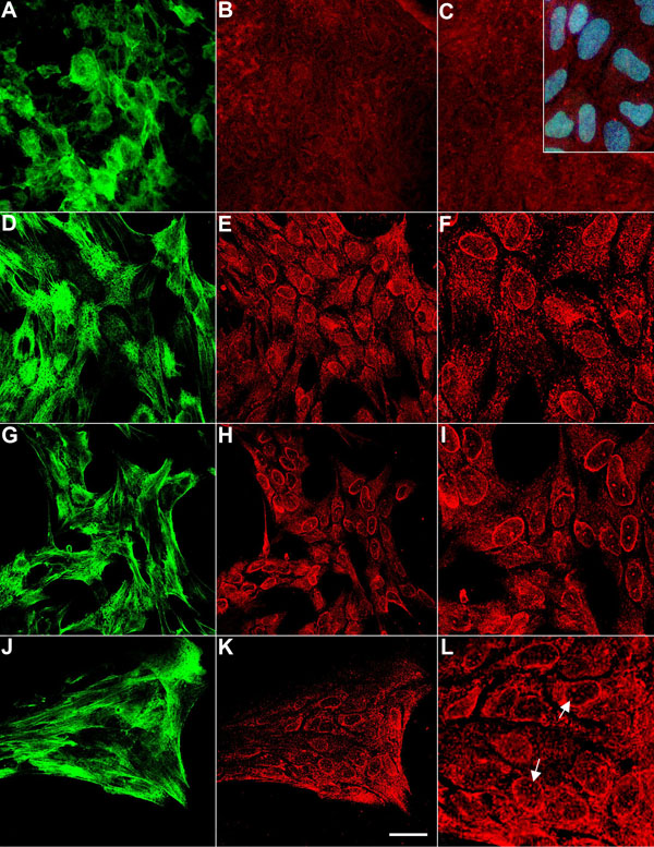
Figure 3.
Localization of phospho-Smad 2/3 in explants. A-C: Explants cultured on laminin. D-F: Explants cultured on vitronectin. G-I: Explants cultured on fibronectin. J-L: explants cultured with 250 pg/ml TGFβ2. A, D, G, J: Localization of α-smooth muscle actin. B, C, E, F, H, I, K, L: Localization of phospho-Smads 2/3. Inset in C: includes Hoechst dye to stain nuclei. Reactivity for phospho-Smads 2/3 was strongly localized in nuclei of cells migrating on vitronectin and fibronectin, whereas little, if any, nuclear localization was detected in cells migrating on laminin. Addition of TGFβ to explants cultured on laminin resulted in characteristic TGFβ-induced changes including the formation of elongated, spindle-shaped cells that showed strong filamentous reactivity for α-smooth muscle actin (J) and nuclear localization of phospho-Smad 2/3 (L, arrows). The scale bar indicates 50 μm in (C, F, I, L) and 100 μm in (A, B, D, E, G, H, J, K).