![]() Figure 2 of
Taliana, Mol Vis 2006;
12:1233-1242.
Figure 2 of
Taliana, Mol Vis 2006;
12:1233-1242.
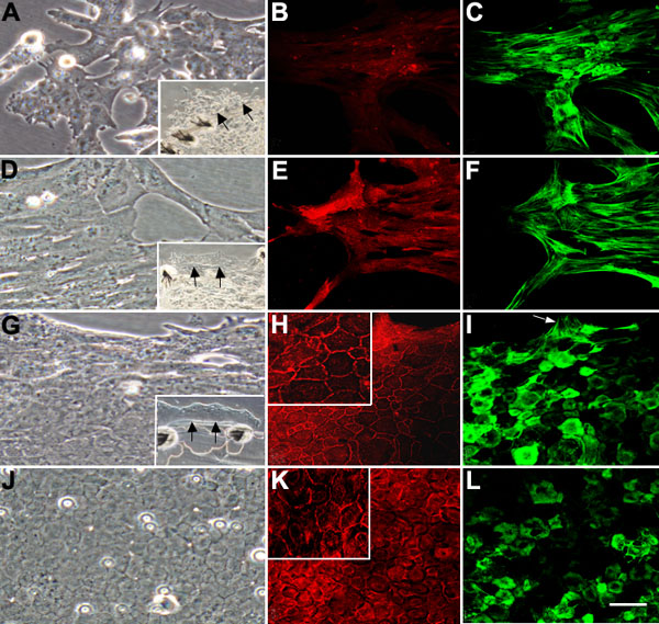
Figure 2.
Characteristics of lens epithelial cell migration in explants cultured on different extracellular matrix substrata. A-C: Explants on vitronectin. D-F: Epithelial cells on fibronectin. G-I: Epithelial cells on laminin. J-L: Standard epithelial explants. A, D, G, J: Phase contrast microscopy. B, E, H, K: ZO-1 localization. C, F, I, L: α-smooth muscle actin localization. A-C: All cells that migrated on vitronectin were elongated and spindle-shaped (typical fibroblastic/myofibroblastic morphology), showed weak, cytoplasmic reactivity for ZO-1 and strong filamentous reactivity for α-smooth muscle actin. D-F: All cells that migrated on fibronectin were elongated and spindle-shaped, showed cytoplasmic reactivity for ZO-1 and strong filamentous reactivity for α-smooth muscle actin. G-I: Most cells that migrated on laminin exhibited an epithelial morphology and strong marginal reactivity for ZO-1 that delineated their cobblestone-like packing arrangement. Many cells showed reactivity for α-smooth muscle actin; except for a few cells at the leading edge (arrow) this reactivity was diffuse and not filamentous. The arrows in the insets in A, D, G indicate the edge of the capsule, beyond which, cells are migrating on the ECM substratum. J- L: Cells on their native substratum, the lens capsule, maintained an epithelial morphology and marginal localization of ZO-1. Many cells also showed diffuse reactivity for α-smooth muscle actin. The scale bar indicates 100 μm.