![]() Figure 4 of
Rozsa, Mol Vis 2006;
12:125-141.
Figure 4 of
Rozsa, Mol Vis 2006;
12:125-141.
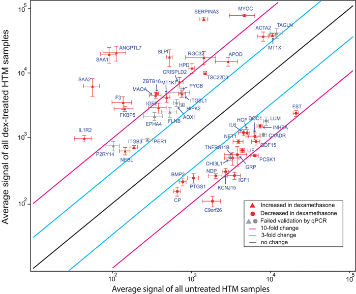
Figure 4. Scatterplot analysis of composite microarray data from three TM cell lines
The logarithmic plot of the composite average GeneChip signal intensities from dexamethasone-treated and untreated cells is shown with error bars representing the standard error of the mean on both axes. Only 52 genes with greater than three fold change in each individual TM cell line are shown. Genes are shown with increased (red triangles) or decreased (red circles) signal intensity in dexamethasone-treated TM cells, relative to untreated cells. Grey symbols indicate genes that failed validation by qPCR (Table 2C). Black, light blue, and pink lines represent fold change boundaries of no change, three fold, and ten fold change, respectively. The Y-axis is the average GeneChip signal intensity from all dexamethasone-treated TM cells; the X-axis is the average GeneChip signal intensity from all untreated TM cells.