![]() Figure 3 of
Rozsa, Mol Vis 2006;
12:125-141.
Figure 3 of
Rozsa, Mol Vis 2006;
12:125-141.
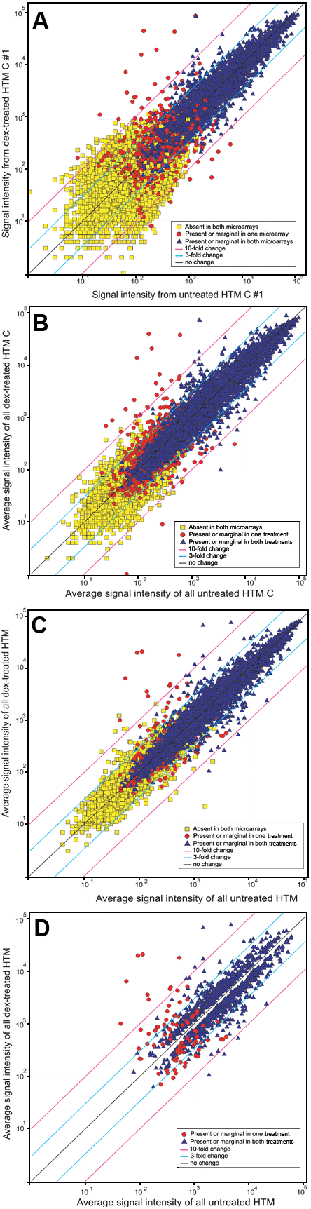
Figure 3. Scatterplots of signal intensities in human trabecular meshwork cells with different treatment
A: Data from one GeneChip from dexamethasone-treated HTM C and data from one GeneChip from untreated HTM C. B: Average signal intensities of data from three GeneChips from three dexamethasone-treated HTM C samples and the average signal intensities of data from three GeneChips from three untreated HTM C samples. C: Average signal intensities of data from nine GeneChips from all dexamethasone-treated TM samples and the average signal intensities of data from 10 GeneChips from all untreated TM samples. D: Data from Panel C after removal of duplicate, nonsignificant, or absent probes as described in Methods. Black, light blue, and pink diagonal lines represent change boundaries of no change, three fold, and ten fold change, respectively.