![]() Figure 2 of
Naskar, Mol Vis 2006;
12:1199-1210.
Figure 2 of
Naskar, Mol Vis 2006;
12:1199-1210.
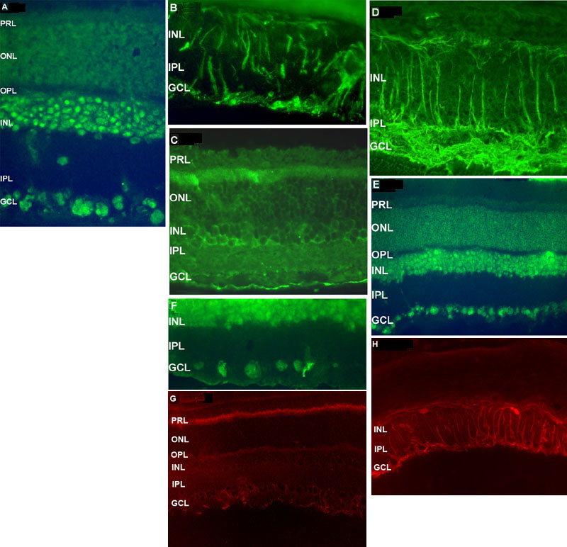
Figure 2. Immunohistochemical detection of c-Myc, GFAP, VEGF, and SA in control and hereditary-intraocular pressure retinas
In the control (A) c-Myc is localized to ganglion cells and the inner nulcear layer (INL, B) In the hereditary-IOP retina c-myc expression is seen in astrocytes and processes of Müller cells. C: In control retinas glial-fibrillary acidic protein (GFAP) is not activated and restricted to the ganglion cell layer (GCL, D) In the hereditary-intraocular pressure retina astrocytes and Müller cells are GFAP positive. E: In the control retina, neurons in the GCL and INL are vascular endothelial growth factor (VEGF) positive. F: In the hereditary-IOP retina, the pattern of VEGF immunoreactivity is unchanged; the number of RGCs are reduced, but still stain for VEGF. G: In control retinas SA is localized to the photoreceptor layer (PRL) whereas H: SA is highly upregulated in the processes of the Müller cells in the hereditary IOP retina. A, B, D, and F were taken at x400 magnification; C, E, G, and H were taken at x200 magnification.