![]() Figure 8 of
Kompella, Mol Vis 2006;
12:1185-1198.
Figure 8 of
Kompella, Mol Vis 2006;
12:1185-1198.
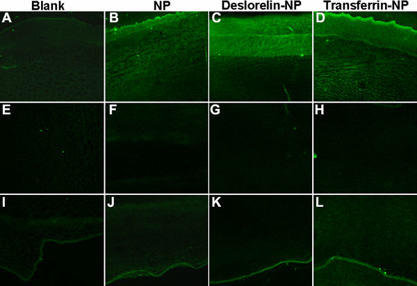
Figure 8.
Representative confocal micrographs to show the uptake of nanoparticles in the epithelial, stromal, and endothelial layers of bovine cornea at the end of 4 h transport study at 37 °C using a modified Ussing chamber setup. A, E, and I represent the top, middle, and lower regions of blank corneal tissue without any nanoparticle exposure. Corresponding regions for NP (B, F, and J), deslorelin-NP (C, G, and K), and transferrin-NP (D, H, and L) are shown in various panels. The confocal images were obtained at a magnification of 20X.